- Image Registration to minimize the effects of patient movement
- Bone Removal to remove the cranium automatically from the dataset
- Deep Learning brain ventricle segmentation to prevent ventricular matter inclusion in quantitative results and improve visual inspection of the maps
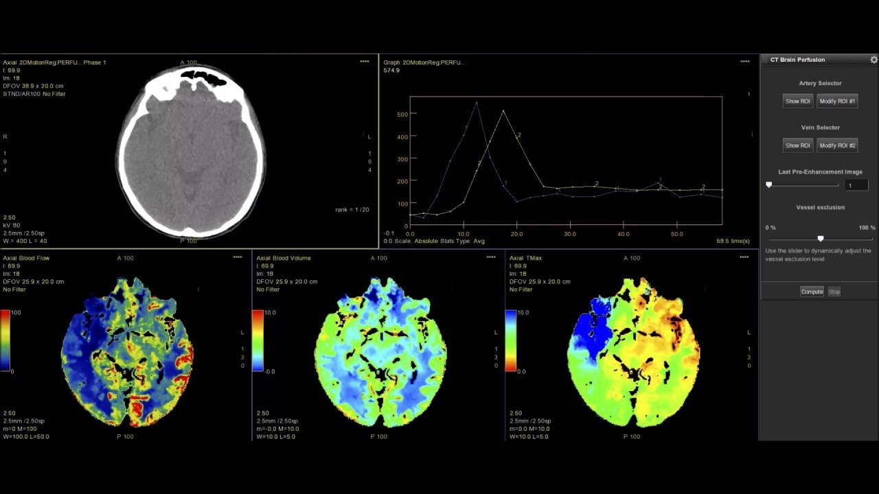
- Automated selection of arterial input and venous output. Both can be easily adjusted if needed.
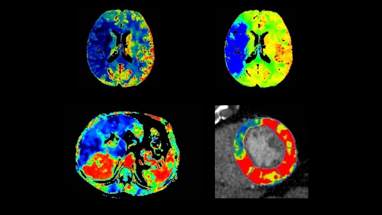
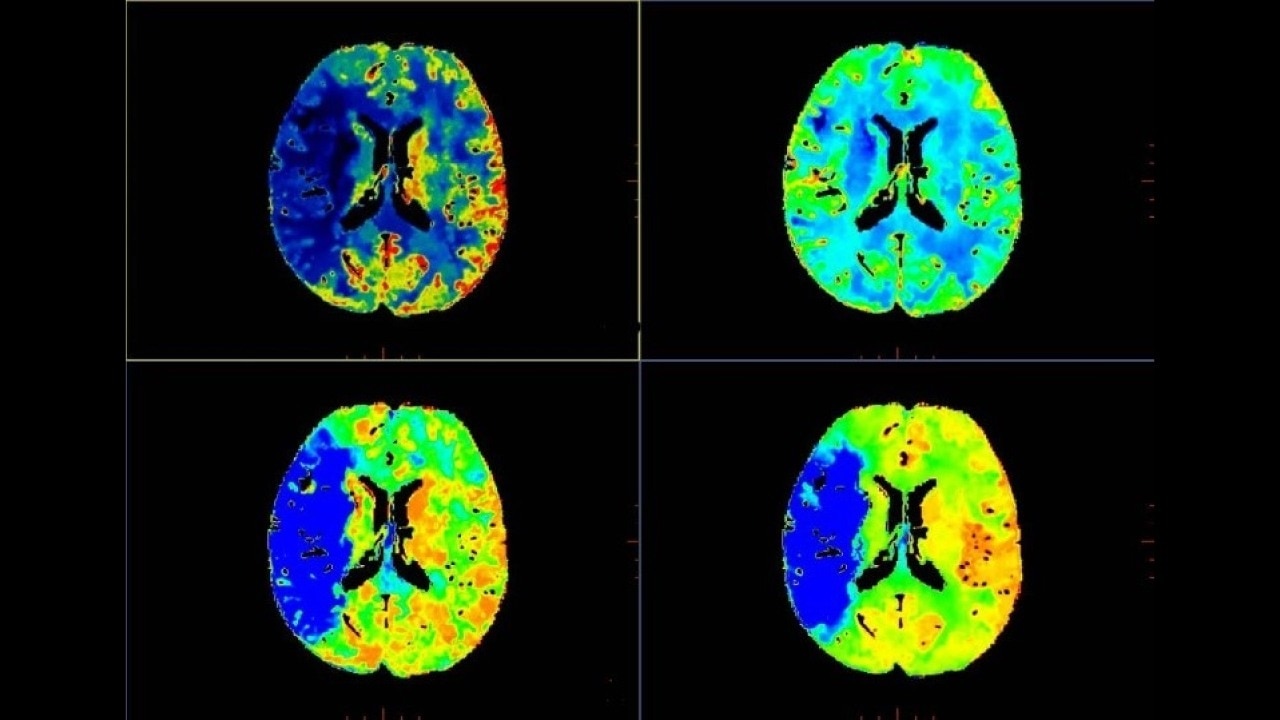
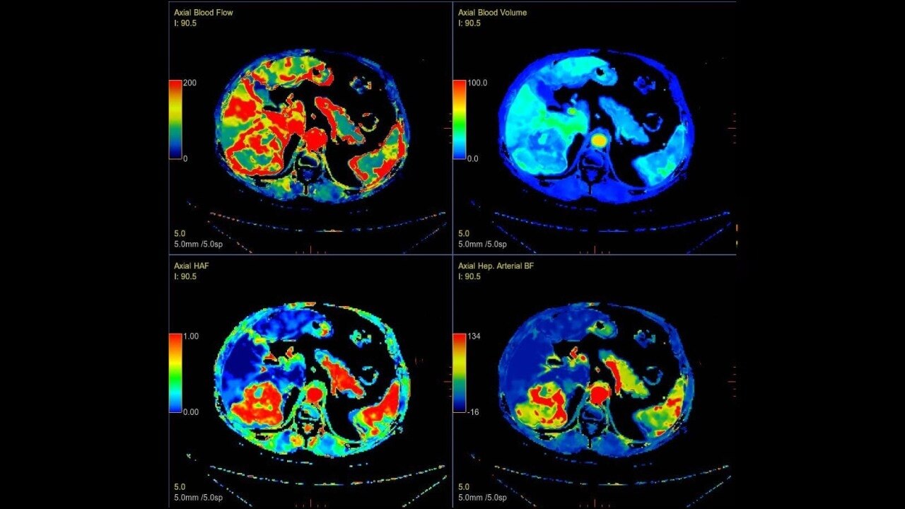
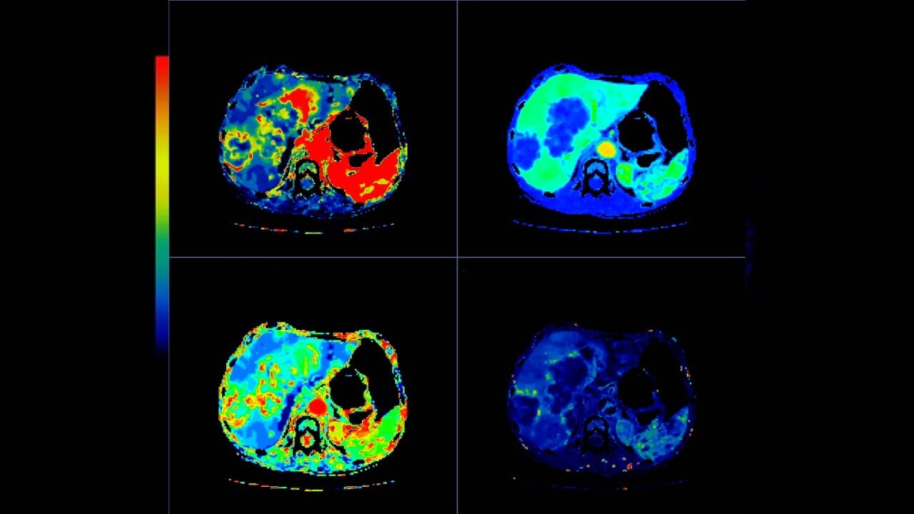
- Automated generation of all functional maps: Blood Flow, Blood Volume, Mean Transit Time, and Transit Time to IRF Peak (Tmax)
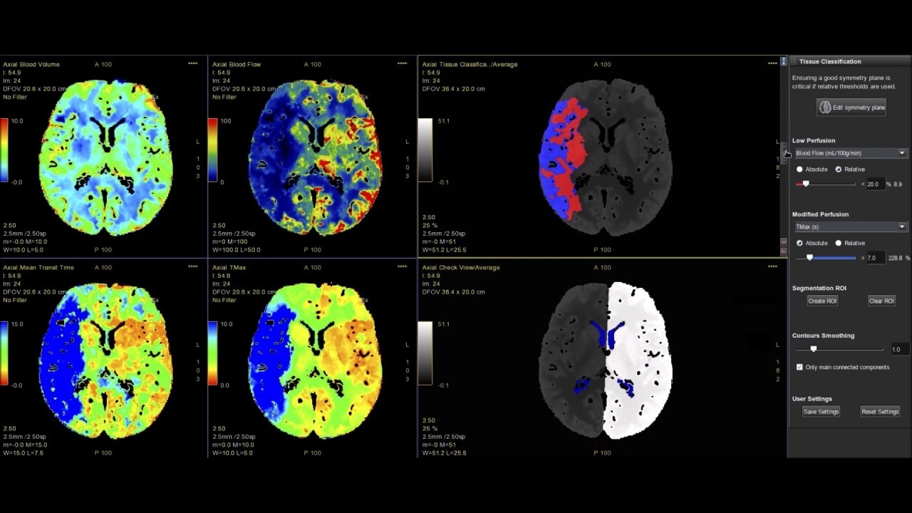
- Automatically define the symmetry plane to be used for mirroring ROIs and relative thresholds
- Tissue Classification enables the visualization of regions that are segmented from absolute or relative values, customizable thresholds and user selectable input maps
- Mismatch volume and ratio are calculated from the modified perfusion volume and low perfusion volume ROIs
Fast, easy-to-use automated software for analyzing CT Perfusion images related to stroke, tumor angiogenesis and dynamic myocardial perfusion.


